Mitochondrial Dynamics & Cellular Response
Mitochondrial Dynamics & Cellular Response
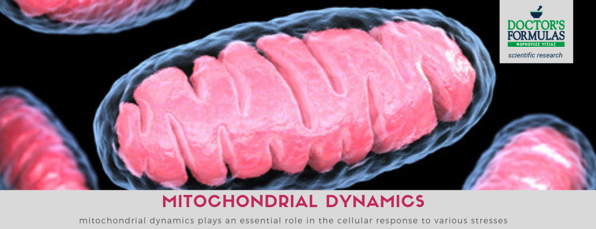
Mitochondria, which have long been seen only as energy producers, are now recognized as the crossroads of many cellular functions. Obviously, they play a key role in producing energy in cells, but they also participate in other processes, such as ion homeostasis, free radical production, and ultimately cell death. Many of their features, such as morphology, cell positioning, proximity to other organelles, are important parameters that need to be taken into account to understand mitochondrial functions.
This is particularly true in adult heart cells where mitochondria, which produce 90% of ATP, occupy 45% of the volume of heart cells and are embedded in a dense and complex organization. These interfaces reflect heart function which requires rhythmic contractions of the pump throughout life and therefore requires a fast and efficient intracellular energy delivery by ATP consumers, cardiomyocytes. In addition, the major phosphorylated metabolites do not change with the increase in work (Balaban, 2012).
This metabolic homeostasis and tight coupling between ATP-consuming mitochondria are two of the cardiac cell specificities and require an optimized cellular organization to ensure efficient energy flow.
Any modification of cellular architecture, but also of the internal organization of mitochondria, could reduce the energy of cells in relation to effects on the cells.
In the majority of cells, mitochondria are able to adapt their morphology and their position according to energy needs and metabolic conditions (Hackenbrock, 1966, Bereiter-Hahn, 1990; Karbowski and Yule, 2003. Rossignol et al., 2004, Mannella, 2006; Benard et al., 2007; Soubannier and McBride, 2009).
This "mitochondrial dynamics" is particularly important during cell division for the quality of mitochondrial control but also plays a role in pathological conditions.
To some extent, the network of mitochondrial morphology is actually the result of several processes, including fusion and fragmentation of organelles that are usually controlled by a complex of protein mechanisms. A linked mitochondrial network is observed in active metabolic cells (Skulachev, 2001), while mitochondria are rather fragmented in dormant cells (Collins et al., 2002). However, it should be borne in mind that mitochondria exhibit high structural and functional specificity with respect to cell functions. For example, in adult cardiac cells, the relationship between mitochondrial morphology and function does not appear to be strict, although cardiomyocytes are metabolically active but exhibit a seemingly fragmented network (Kuznetsov et al., 2009).
Mitochondrial dynamics, however, depends on the cellular environment and its limitations.
In adult cardiomyocytes, the large number of myonomatics, the presence of a rigid cell skeleton and the dense mitochondrial network clearly hamper mitochondrial movements (Vendelín et al., 2005). In addition, the arrangement of the various organelles is so critical to the cardiac cell function that mitochondrial morphology needs to be effectively controlled (Wilding et al., 2006; Piquereau et al., 2010).
Mitochondrial disruption is recognized as a critical event in a number of pathological conditions, including hypoxia, ischemic lesions, strokes, diabetes mellitus, The carnitine acyltransferase pathway has been shown to be critical for maintaining the normal functioning of mitochondria. Fatty acids are transported via carnitine to mitochondria for subsequent oxidation to eventually produce ATP.
Carnitine is necessary because increasing levels of free fatty acids can induce mitochondrial dysfunction resulting in cell death and / or increased secondary production of active forms of oxygen.
Studies have also shown that carnitine has a protective effect on both mitochondria and the whole cell by inhibiting the damage caused by free fatty acids in the mitochondrial membrane and consequently its secondary effects. The protective effects of carnitine and its related metabolites have been documented to stem from the improvement of energy metabolism and the inhibition of electron leakage by mitochondrial electron transport systems.
Carnitine has also been shown to reduce free fatty acids in serum and tissues and prevent tissue damage.
Mitochondria are also important signaling transducers in the apoptotic pathway in a variety of disease states. Previous studies have shown a close relationship between carnitine and mitochondria. Cultured cardiac myocytes were studied to evaluate mitochondrial changes during apoptosis. Carnitine has been shown to inhibit mitochondrial-dependent apoptosis both in vivo and in vitro.
The role of mitochondria in cardiovascular pathologies has been extensively tested. In cardiovascular diseases, there is increased production of active oxygen species (ROS). This leads to endothelial dysfunction and disorder of endothelium-dependent vasodilatation, thereby reducing the bioavailability of NO. Studies on the mechanism underpinning the effects of carnitine on cardiovascular disease have shown that chronic administration of carnitine can reduce blood pressure and mitigate the inflammatory process associated with arterial hypertension.
Carnitine therapy has also been shown to increase post-ischemic blood flow in patients with peripheral vascular disease, indicative of improvement in functional circulation.
Carnitine has been shown to reduce ischemia-reperfusion injury by preventing the accumulation of long-chain acetyl-CoA and subsequent production of ROS from damaged mitochondria. It also improves repair mechanisms for the oxidative damage caused to the phospholipid membrane and reduces ischemia-induced apoptosis and subsequent remodeling of the left ventricle.
Carnitine also restores endothelial dysfunction present in hypertensive and has the potential to increase the release of vasodilator agents. The production and availability of NO play an important role in determining the functions of endothelial cells. Reductions in the expression of endothelial nitric oxide synthase (eNOS) and inactivity leading to reduced bioavailable NO production have been associated with endothelial dysfunction and vascular remodeling. In cardiomyocytes, mitochondria are abundant (as much as 45% of the cell volume) to fulfill the dominant cell dependence on mitochondria to produce ATP to support contractile function and cardiac output. Thus the mitochondria are densely localized in a linear configuration. In addition, mitochondria are the major source of peroxide in cardiomyocytes, particularly in response to reduced oxygen availability (Hool et al., 2005). This has led to the development of treatments for ischemia-reperfusion injury using agents such as CoQ10, which serves multiple molecular roles in mitochondria (Ebaddy, 2001).
CoQ10 improves contractile function while preventing calcium overload and preserves diastolic function.
A more recent study showed that CoQ10 treatment improved diastolic function during reperfusion while maintaining higher levels of ATP, coronary vasodilation with nitrite and increased coronary flow (Whitman et al., 1997).
CoQ10 also specifically binds to a site in the internal mitochondrial membrane that inhibits the mitochondrial permeability transition. This is a large channel of conduction, which when opened can activate the collapse of mitochondrial strength and membrane potential. In cell death, this signaling pathway leads to the breakdown of homeostasis ions and oxidative phosphorylation, particularly after ischemia and reperfusion. CoQ10 protects creatine kinase and other essential proteins from oxidative inactivation during reperfusion, a function that is important for maintaining energy metabolism and cardiac output. Ferrara et al. (1995) demonstrated that chronic CoQ10 treatment in models protects against heart disease due to oxidative stress.
CoQ10 can inhibit lipid peroxidation in mitochondria and oxidation of proteins and DNA.
It plays an important role in the mitochondrial respiratory chain for the production of ATP, in the extra-mitochondrial transfer of electrons as well as in the regulation of NADH oxidoreductase activity in the plasma membrane and also exerts a dynamic redox action on both Golgi and lysosomes. There is another possible molecular effect of CoQ10, related to the ability of CoQ10 to regulate and change genomic expression. CoQ10 has been shown to target expression in multiple genes, particularly those involved in cellular signaling and intermediate metabolism.
Many of these genes include those governing nucleic and other enzymes, transcription factors, muscle fiber components, growth factors, receptors and signal transduction of the activated receptor and other metabolic pathways. Thus, the metabolic flux regulation and control gene can explain many of the cardiovascular and other effects of CoQ10 according to which it can act advantageously at multiple sites in the pathophysiological sequence.
At the clinical level, protection against myocardial ischemia has been demonstrated in two double-blind, placebo-controlled trials of CoQ10 in patients with ischemic heart disease with decreased angina, with improved exercise resistance and reduced ECG ischemic changes. These beneficial effects may be related to increased efficacy of mitochondrial energy production after the heart attack, as has been shown in patients with ischemic heart disease.
Taurine is a ubiquitous compound, found in very high concentrations in the heart and muscles. Although taurine is classified as an amino acid, it does not participate in the formation of peptide bonds. However, the amino group of taurine has been implicated in a series of significant coupling reactions as well as hypochlorite acid scavenging.
Because taurine is a fairly inactive compound, it is an ideal regulator of basic procedures, such as osmotic pressure, cation homeostasis, enzyme activity, receptor regulation, cell growth, and cell signaling.
The depletion of taurine leads to the development of myocardial problems and shows a role for taurine in maintaining normal contractile function. This function of taurine is caused by changes in the activity of the key carriers of Ca2 + and by the formation of myofibrillar sensitivity. Taurine stabilizes the membranes by direct interactions with the phospholipids. It also serves as a modulator of protein kinases and phosphatases within the cardiomyocytes.
References:
-
Mitochondrial dynamics in the adult cardiomyocytes: Which roles for a highly specialized cell?Article (PDF Available) in Frontiers in Physiology 4:102 · May 2013 with 295 ReadsDOI: 10.3389/fphys.2013.00102 · Source: PubMed
-
The role of mitochondrial function and cellular bioenergetics in ageing and disease. M.D. Brand, A.L. Orr, I.V. Perevoshchikova, and C.L. Quinlan. doi: 10.1111/bjd.12208
-
Mitochondrial energetics in the kidney. Pallavi Bhargava and Rick G. Schnellmann. Published online 2017 Aug 14. doi: 10.1038/nrneph.2017.107
-
Coenzyme Q10 in cardiovascular disease. Salvatore Pepe a,b,c, Silvana F. Marasco a,b,c, Steven J. Haas b, Freya L. Sheeran b,c, Henry Krum b, Franklin L. Rosenfeldt a,b,c,* https://pdfs.semanticscholar.org/f4f4/1461240f5f11abe0f338ca0535c974649da8.pdf
-
Targeting mitochondrial dysfunction and oxidative stress in heart failure: challenges and opportunities. Ligia Akemi Kiyuna,1 Rudá Prestes e Albuquerque,1 Che-Hong Chen,2 Daria Mochly-Rosen,2 and Julio Cesar Batista Ferreira1,# Published online 2018 Sep 15. doi: 10.1016/j.freeradbiomed.2018.09.019