The important role of vitamin K beyond clotting
The different roles and actions of K1 and K2, isoforms of vitamin K
Vitamin K has been known for more than 80 years for its role in blood clotting. It was first recognized in 1936 as a key factor involved in blood clotting. When the chickens were fed a low fat diet, they had a significantly lower clotting capacity, resulting in severe bleeding.
The lipid fraction of the diet was analyzed and a new anti-haemorrhagic agent was discovered. This fat soluble agent was given the letter K from the German word "Koagulation" and was considered to be only necessary for its anti-haemorrhagic characteristic. Since then, non-coagulant functions have been discovered and have attracted research interest in various fields around the world.
The vitamin K family consists mainly of two vitamins:
vitamin K1 (also known as phylloquinone) and
vitamin K2 (also called menakinone).
The discovery of the various isoforms of vitamin K begins to clarify the important role of vitamin K beyond clotting.
The functions of K2 prove to be beneficial in terms of both cardiovascular risk and bone metabolism. There is a growing body of evidence suggesting that vitamin K2 is involved in multiple cellular processes and may play a protective role in various organs throughout the human body.
This study highlights and summarizes the differences between the intake and function of vitamin K1 and vitamin K2. In exploring the non-clotting but extracellular activity of vitamin K, it is clear that K2 in its various forms is the major point of difference for this activity. Therefore, although history and nomenclature have classified K1 and K2 into the same category, these molecules have a very different effect on the body.
Vitamin K1 is the predominant form of vitamin K present in the diet. Vitamin K1 is found mainly in green vegetables and chlorophyll in plants, while menacinones K2 are composed of bacteria and are found primarily in foods where bacteria form part of the production process.
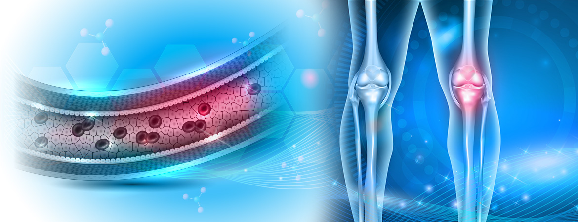
The most well-known function of Vitamin K is as a co-factor for activating vitamin K-dependent coagulation factors. Through the post-translational modification of glutamate (Glu) residues into coagulation factors, vitamin K enables post-translational carboxylation, which allows high affinity binding to calcium support in negatively charged regions of the phospholipid membrane and the haemostasis.
Vitamins K1 and K2 can act as co-factors in the process of carboxylation of vitamin K-dependent proteins (VKDPs). With this in mind, vitamin K serves as a cofactor of γ-glutamyl carboxylase (GGCX), which catalyzes the glutamate (Glu) residues of vitamin K-dependent proteins (VKDPs) on γ-carboxyl glutamyl ). This process is guided by the oxidation of Vitamin K-Hydroquinone (KH2) to Epoxy-Vitamin K (KO) in the Vitamin K. The Vitamin K-Oxidoreductase (VKOR) converts Epoxide-Vitamin K (KO) back to vitamin K-hydroquinone (KH2), creating a process of recycling vitamin K.
Vitamin K-dependent proteins (VKDPs) are categorized as hepatic and extracellular vitamin K-dependent proteins (VKDPs).
Vitamin K-dependent hepatic proteins (VKDPs) include coagulation factors II, VII, IX, X and anticoagulant protein C, protein S and protein Z, which are involved in regulating blood coagulation. Vitamin K-dependent extracellular proteins (VKDPs) include the Gla Protein (MGP), osteocalcin and Gla rich protein (GRP).
These vitamin K-dependent proteins (VKDPs) are primarily involved in maintaining bone homeostasis as well as in inhibiting ectopic calcification.
Basal Gla Protein (MGP) is mainly expressed in vascular smooth muscle cells and chondrocytes. This protein is known to inhibit extracellular mineralization of vascular lesions and is involved in vascular remodeling.
Osteocalcin can be designated as a bone protein specific protein and is involved in the regulation of metal deposition. Gla-rich protein (GRP) can be found in calcified cartilage and in the vascular system, where it directly binds and inhibits the formation / maturation of crystals and calcification of vascular smooth muscle cells.
Osteocalcin can be designated as a bone protein specific protein and is involved in the regulation of metal deposition. Gla-rich protein (GRP) can be found in calcified cartilage and in the vascular system, where it directly binds and inhibits the formation / maturation of crystals and calcification of vascular smooth muscle cells.
In addition to the role of vitamin K in carboxylation of vitamin K-dependent proteins (VKDPs), recent studies have suggested a role for vitamin K as an antioxidant.
The enzyme VKORC1, called VKORC1-like 1 (VKORC1L1), is expressed in many tissues. VKORC1L1 has been shown to mediate vitamin K-dependent intracellular antioxidant function in human cell membranes. Where it protects cell membranes from lipid peroxidation.
This antioxidant effect is reduced by the presence of warfarin. Both vitamin K1 and K2 inhibit oxidative stress in neuronal cells and primary oligodendrocytes through inhibition of 12-lipoxygenase. In addition to its antioxidant properties, it has been reported that vitamin K facilitates ATP production and rescues mitochondrial dysfunction.
Of all the menacinones, MK-7 is more efficiently absorbed and has the highest bioavailability.
All forms of vitamin K are absorbed by the enterocytes into the small intestine and are packaged in chylomicrons during absorption. These chylomicrons are then absorbed by the liver. Vitamin K1 is preferably preserved in the liver to aid in the carboxylation of coagulation factors.
In contrast, Vitamin K2, especially long-chain derivatives, is redistributed into the circulation and is available for extrapulmonary tissues such as bones and vessels.
Vitamin K2-dependent proteins are variously expressed in both soft and hard tissues. The role of K1 in coagulation has been established. While the position of vitamin K2 in health and disease is well recognized in the context of cardiovascular disease, bone and fracture development, chronic kidney disease and additional evidence, further evidence of vitamin K2 roles liver disease, immune function, neurological diseases and obesity.
Vascular calcification is an active process that causes cardiovascular disease, the largest killer in the world. It is known that vitamin K2-dependent proteins activate a protective mechanism that inhibits the development of vascular calcification.
In addition, vitamin K2, in the form of MK-7, has been shown in numerous studies in healthy and patients that it has a long-term protective effect on the development of calcification. In addition, several studies have shown an overall reduction in the risk of cardiovascular disease. Even a regression of arterial stiffness and improved vascular elasticity was observed in healthy population groups after supplementation.
Interestingly, in a study investigating all isoforms of vitamins K1 and K2, only K2 was effective and beneficial to cardiovascular health and not K1. In addition, the role of K2 in cardiovascular disease has been evident and has been the subject of many large clinical trials performed worldwide supplementing vitamin K2 in a variety of cardiovascular patients, the results of which will add more to the role of vitamin K2 in the heart.
Fractures and bone quality are important for the elderly population. Vitamin K2 is known to improve bone quality, which in turn reduces the risk of fracture, as demonstrated by numerous studies with population groups over 50 years of age. In addition, there are ongoing population studies that will further highlight the role of vitamin K2 in bone growth, health and maintenance.
Long-term supplementation with vitamin K2 has been shown to reduce the risk of developing diabetes. The largest study, with 38,000 men and women 20-70 years old, showed that only 10 µg / day of K2 reduces the risk of diabetes by 7%.
The mechanism by which K2 can act begins to unfold ...
Vitamin K2 activates osteocalcin, which has been shown in vitro to promote pancreatic beta cell proliferation as well as to increase insulin production and Cyclin D1 expression. This mechanism is under investigation and it is assumed that osteocalcin, lectin and adiponectin have a complex network for glucose metabolism that can be regulated by vitamin K2.
The role of vitamin K in the liver has been well established in the production of coagulation factors and activation of vitamin K-dependent proteins (VKDPs). While the majority of research has focused on K1, K2 has higher bioactivity and can act similarly on liver tissue. In addition, emerging research on vitamin K2 demonstrates a regenerative effect on ovarian cells, as well as maturation of hepatocytes in stem cell cultures. This suggests that there may be developmental importance of K2 in the liver.
The status of the basic dephosphorylated-non-carboxylated-protein GLA (dp-ucMGP) is an accepted research indicator for vitamin K deficiency that was first described in patients with chronic renal disease. The dephosphorylated non-carboxylated-basic protein GLA (dp-ucMGP) is associated with the progression of chronic renal disease, as patients with chronic renal disease subsequently have higher levels of circulating dephosphorylated carboxylphosphoryl fundamental substance (dp-ucMGP).
Vitamin K2 supplementation has been shown to improve renal artery function and prevent further development of renal artery calcification.
This has overall benefit for kidney function. In addition, vitamin K2 supplementation has been shown to improve glomerular filtration in a group of patients. Vitamin K2 in chronic kidney disease research is high and there are many large-scale studies in progress.
In recent years, ex vivo studies have shown a previously unknown immunomodulatory role for vitamin K2.
First, it was shown that MK-7 regulates the expression of TNF-α, IL-1α and IL-1β. In line with this finding, K2 reduces T-cell proliferation in healthy individuals, whereas vitamin K1 did not. This has been further substantiated with T-cells from a larger number of children with atopic dermatitis and healthy controls, as well as a separate study with dialysis patients. Both of these studies showed that K2 reduced the number of activated T-cells and their proliferation.
Thus, the accumulated evidence points to a new role of vitamin K2 as an immunosuppressive agent. This needs to be further developed by then, a new physiological mechanism by which vitamin K2 may aid immune modulation may be hypothesized, although it requires further study.
The extra-hepatic effect of vitamin K2 has been elucidated in vivo by altering the activity of K2 recycling enzymes in different tissues. Apparently, a subset of enzymes of vitamin K2 has been shown to be highly expressed in the brain. A protective effect of K2 on neurons in vitro has been documented.
MK-4 improved energy production and rescued the PINK1 mutation found in Parkinson's disease. More recently, research has determined the protection of neurons from vitamin K2 through a novel mechanism involving the P38 MAP kinase pathway. Furthermore, various analogues of K2 have been found to be crucial in neuronal differentiation.
The first epidemiological study of K2 on neuronal activity included a group of 45 MS patients and 29 healthy volunteers. K2 levels were significantly reduced in patients with multiple sclerosis compared to controls dependent on gender and age.
Also, K2 levels were associated with neurological convulsions and optic nerve lesions. These emerging studies suggest a potentially important role for vitamin K2 in neurological development and disease.
The connection between osteocalcin and adiponectin has been strongly suggested. However, the way in which any such action takes place remains unclear. The immediate increase in non-carboxylated osteocalcin (ucOC) levels reduced fat mass and improved glucose metabolism in models. Further, a mechanism has been implicated by Ding et al., Where it was found that VKORC1L1 promotes lipogenesis.
Consequently, down-regulation of VKORC1L1 upregulation increased intracellular K2 levels and inhibited proliferation. Studies of human cohorts have shown improvements in body weight, waist circumference, body composition, visceral fat and diabetes mellitus from supplementation with K2.
All of these indicate an overall beneficial effect of vitamin K2 on glucose and fat metabolism, which requires further research to confirm.
Story Source: International Journal Molecular Sciences 2019